Pelvic Anatomy : Pelvic Contents Female. Using a speculum, a doctor can examine the vulva, vagina, and cervix.the strength of the pelvic muscles can also be tested. Describe the boundaries and subdivisions of the pelvis. The right and left hip bones also converge anteriorly to attach to each other. The pelvis is a bony ring, or circle structure. The superior two thirds correspond to the uterine body and the inferior third to the cervix.
Use the mouse scroll wheel to move the images up and down alternatively use the tiny arrows (>>) on both side of the image to move the images.>>) on both side of the image to move the images. The superior two thirds correspond to the uterine body and the inferior third to the cervix. 100 % 0 % videos / pods. The lumbosacral plexus is formed by the lumbosacral trunk and the ventral rami of the first to third sacral nerves, and part of the fourth sacral nerve. Surgical anatomy of the female pelvis by laparoscopy.
Application Of Pelvic Binders By Student Paramedics An Observational Cohort Study Journal Of Paramedic Practice from www.paramedicpractice.com The pelvis's frame is made up of the bones of the pelvis, which connect the axial skeleton to the femurs, and therefore acts in weight bearing of the upper body. Gross anatomy of the pelvis—namely the bladder, uterus, fallopian tubes, ovaries, rectum, and the muscles—has remained unchanged; Each innominate bone is composed of three fused bones: This mri male pelvis axial cross sectional anatomy tool is absolutely free to use. Use the mouse scroll wheel to move the images up and down alternatively use the tiny arrows (>>) on both side of the image to move the images.>>) on both side of the image to move the images. At the top of this bony ring, if you were to put your hands on your hips, you wouldn. Anatomy of female pelvic area facebook twitter linkedin pinterest print fertility and reproductive health pelvic floor disorders fertility, pregnancy and childbirth women's health. The lining of the uterus.
It is strengthened and supported by several joints and ligaments.
• pelvis begins at the iliac crests and ends at the symphysis pubis. Each innominate bone is composed of three fused bones: Describe the anatomy of the pelvic wall, bones, joints & muscles. The pelvis is inferior most part of the trunk. Sometimes we call all of the structures within this ring the pelvic girdle, which can also sometimes include the hips and the abdomen (having pain somewhere around your pelvic girdle? Understanding what each term refers to and its purpose can help you make the wisest decision. Ct body (lymph nodes) ct. {{configctrl2.info.metadescription}} this site uses cookies. • divided into the true and false pelvis by the iliopectineal line. The bony pelvis consists of the two hip bones (also known as innominate or pelvic bones), the sacrum and the coccyx. Consisting of the pelvic girdle and perineum, it supports the urinary and reproductive organs. The male pelvis is different from a female's. There are four articulations within the pelvis:
• divided into the true and false pelvis by the iliopectineal line. The uterus represents the essential landmark of pelvic anatomy. It is usually divided into two separate anatomic regions: The term `pelvis` can refer to the pelvic skeleton (also known as the pelvic girdle), which is the skeleton embedded in the lower part of the trunk, connecting the axial skeleton to the lower extremities. However, knowledge of the anatomy of various structures that surround these organs has evolved over time.
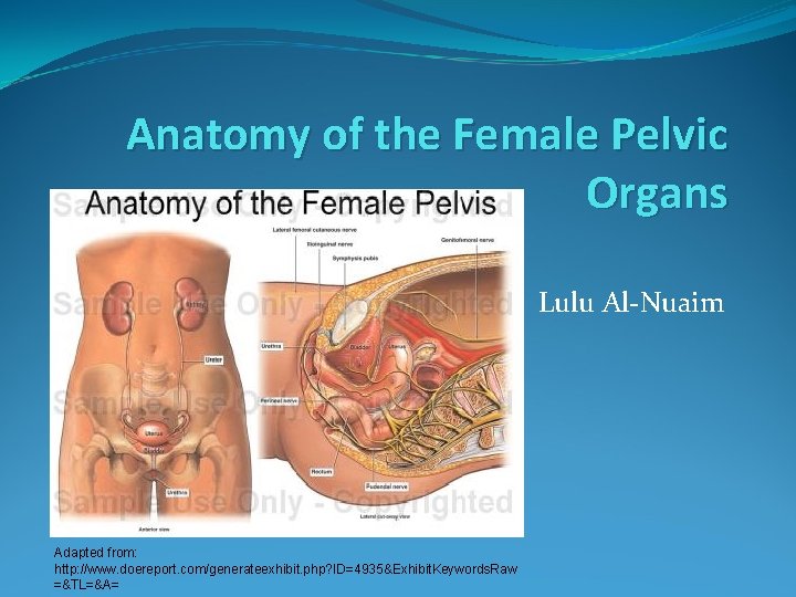
Anatomy Of The Female Pelvic Organs Lulu Alnuaim from slidetodoc.com Check out these posts for help!). Gross anatomy of the pelvis—namely the bladder, uterus, fallopian tubes, ovaries, rectum, and the muscles—has remained unchanged; The male pelvic floor is a complex structure made up of muscles, ligaments, nerves and fascia. The pelvic girdle and pelvic spine. It is based on a 3d scan. It's located between the abdomen and the legs. The pelvis's frame is made up of the bones of the pelvis, which connect the axial skeleton to the femurs, and therefore acts in weight bearing of the upper body. The lumbosacral plexus is formed by the lumbosacral trunk and the ventral rami of the first to third sacral nerves, and part of the fourth sacral nerve.
The pelvis is a basin shaped bony structure formed by the combination of two pelvic bones (hip bones or innominate bones) and the sacrum.
Chris battista 0 % topic. Describe the components & function of the pelvic diaphragm. The right and left hip bones also converge anteriorly to attach to each other. The male pelvis is different from a female's. The pelvic girdle (hip girdle) is formed by a single bone, the hip bone or coxal bone (coxal = hip), which serves as the attachment point for each lower limb. Anatomy of female pelvic area facebook twitter linkedin pinterest print fertility and reproductive health pelvic floor disorders fertility, pregnancy and childbirth women's health. • divided into the true and false pelvis by the iliopectineal line. It is located in the middle of the pelvis between the urinary bladder lying before and the large bowel lying behind it. Each innominate bone is composed of three fused bones: Consisting of the pelvic girdle and perineum, it supports the urinary and reproductive organs. The pelvic bones and the sacrum. It is usually divided into two separate anatomic regions: The nerves of the pelvis include:
A 3d rotatable model of the bony structures of the pelvis: It is usually divided into two separate anatomic regions: Laparoscopic anatomy of the female pelvic region. The uterus, sometimes called the womb, is a muscular organ located in the pelvis. The pelvis's frame is made up of the bones of the pelvis, which connect the axial skeleton to the femurs, and therefore acts in weight bearing of the upper body.
Pelvic Anatomy Clinic Prints from static.wixstatic.com It is usually divided into two separate anatomic regions: The pelvic bones and the sacrum. The pelvic girdle and pelvic spine. The pelvis is the part of the body located between the abdomen and the thighs. Pelvic ring formed from 2 innominate bones. The pelvis (plural pelves or pelvises) is either the lower part of the trunk of the human body between the abdomen and the thighs (sometimes also called pelvic region of the trunk) or the skeleton embedded in it (sometimes also called bony pelvis, or pelvic skeleton). Articulate posteriorly with the sacrum and anteriorly through pubis symphysis; Surgical anatomy of the female pelvis by laparoscopy.
At the top of this bony ring, if you were to put your hands on your hips, you wouldn.
The anatomy of the pelvis varies depending on whether you are male or female. The pelvis is a basin shaped bony structure formed by the combination of two pelvic bones (hip bones or innominate bones) and the sacrum. Describe the components & function of the pelvic diaphragm. Anatomy of female pelvic area facebook twitter linkedin pinterest print fertility and reproductive health pelvic floor disorders fertility, pregnancy and childbirth women's health. It provides attachment to some important muscles in the region, and forms a cavity which accommodates several important internal organs. Differentiate the different types of the female pelvis. • pelvis begins at the iliac crests and ends at the symphysis pubis. The superior two thirds correspond to the uterine body and the inferior third to the cervix. Let's focus in on the pelvic anatomy: {{configctrl2.info.metadescription}} this site uses cookies. Gross anatomy of the pelvis—namely the bladder, uterus, fallopian tubes, ovaries, rectum, and the muscles—has remained unchanged; The bony pelvis consists of the two hip bones (also known as innominate or pelvic bones), the sacrum and the coccyx. Ct body (lymph nodes) ct.